Monkey next to negative photos of serial sections of the myosin heavy chain profile of a monkey chewing muscle

Chewing Muscle Fiber Architecture

We study scaling relationships of jaw-muscle architecture and musculoskeletal correlates of the primate feeding system and feeding behavior/diet.

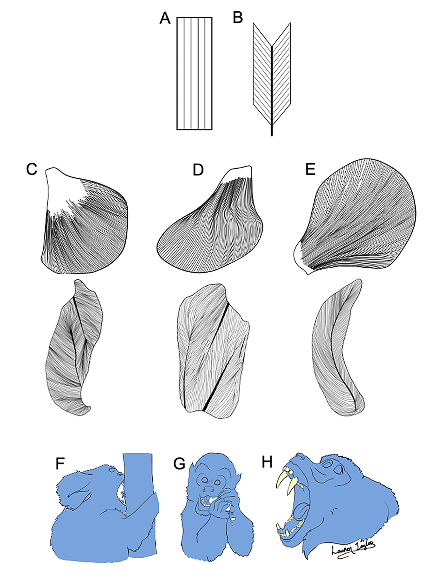
Primates face ecological challenges associated with gaining access to and processing available food resources. Gape (mouth opening) and bite force are key performance variables, and these two performances cannot be simultaneously maximized without modifying other aspects of the masticatory apparatus. Our architectural studies of the jaw-closing muscles are focused on how animals meet these competing demands.

We have been exploring how key determinants of muscle stretch (fiber length) and muscle force (physiological cross-sectional area, PCSA), scale with body and skull size and muscle leverage and testing hypotheses of jaw-muscle architecture, function and evolution in relation to both dietary variation and social signaling and aggressive biting behaviors. In collaboration with PIs Dr. Janine Chalk-Wilayto (Mercer University), Dr. Megan Holmes (Duke University), Dr. Myra Laird (University of Pennsylvania), and Dr. Claire Terhune (University of Arkansas), we are also testing hypotheses about how jaw-adductor fiber architecture changes during ontogeny in the hard-object feeding tufted capuchin monkey. View Dr. Taylor's ResearchGate page for publications resulting from this work.

Top: Skeletal muscle fibers may be oriented in parallel (A) or at an angle (B) relative to the axis of force generation. For muscles of comparable volume, parallel-fibered muscles pack in fewer, but longer fibers, making them well-suited for producing large excursions, whereas pinnate-fibered muscles pack in shorter, but more fibers, and thus are designed for producing large muscle forces. Middle: The masseter (C) and medial pterygoid (D) are multipinnate (i.e., fibers oriented in multiple directions), whereas the temporalis (E) is bipinnate (i.e., fibers oriented in two directions). Upper row represents whole muscles, lower row represents corresponding muscles in cross-section. All are left-sided muscles. Bottom: Fiber length and orientation have significant functional implications for muscle stretch/gape and muscle and bite force related to feeding (F, G) and for gape behaviors unrelated to feeding such as wide-mouth display (H).

In this image:

Top: Skeletal muscle fibers may be oriented in parallel (A) or at an angle (B) relative to the axis of force generation. For muscles of comparable volume, parallel-fibered muscles pack in fewer, but longer fibers, making them well-suited for producing large excursions, whereas pinnate-fibered muscles pack in shorter, but more fibers, and thus are designed for producing large muscle forces. Middle: The masseter (C) and medial pterygoid (D) are multipinnate (i.e., fibers oriented in multiple directions), whereas the temporalis (E) is bipinnate (i.e., fibers oriented in two directions). Upper row represents whole muscles, lower row represents corresponding muscles in cross-section. All are left-sided muscles. Bottom: Fiber length and orientation have significant functional implications for muscle stretch/gape and muscle and bite force related to feeding (F, G) and for gape behaviors unrelated to feeding such as wide-mouth display (H).